

Back or neck symptoms do not always remain in one place. Pain may affect several areas, tingling may travel into an arm or leg, or a healthcare professional may need to assess more than one part of the spine.
In these situations, a full spine MRI may provide detailed images of the cervical, thoracic and lumbar spine during one examination.
However, scanning the entire spine is not automatically better than examining one specific region. The most appropriate scan depends on your symptoms, medical history and the clinical question that needs answering.
This guide explains what a full spine MRI shows, when it may be considered and how it differs from a targeted spine or full-body MRI, so you can have a more informed conversation about the next step.
What is a full spine MRI?
A full spine MRI, sometimes called a whole spine MRI, is a non-invasive examination covering the three main regions of the spine:
| Spine region | Area examined |
| Cervical spine | The neck and upper part of the spine |
| Thoracic spine | The middle of the back |
| Lumbar spine | The lower back |
MRI uses a strong magnetic field and radio waves to create detailed images. It does not use the ionising radiation associated with X-rays and CT scans.
An MRI of the spine can show the vertebrae, intervertebral discs, spinal canal, spinal cord, nerve roots, joints, ligaments and surrounding soft tissues.
VitalScan provides a private full spine MRI scan covering all three spinal regions at its Milton Keynes clinic.
What does a full spine MRI show?
A full spine MRI can reveal structural changes affecting one or several parts of the spine.
| Structure | What the MRI may help assess |
| Vertebrae | Alignment, fractures, bone marrow changes and other structural abnormalities |
| Intervertebral discs | Degeneration, loss of disc height, bulging or herniation |
| Spinal canal | Areas of narrowing, also known as spinal stenosis |
| Spinal cord | Compression, inflammation or other abnormalities |
| Nerve roots | Narrowing or compression where nerves leave the spine |
| Joints and ligaments | Arthritis, thickening, injury or degenerative changes |
| Surrounding tissues | Inflammation, infection or unusual masses requiring further investigation |
An MRI scan for the spinal cord can be particularly useful when symptoms include weakness, numbness, balance changes or difficulty walking. Because the spinal cord and nerves carry messages throughout the body, symptoms may appear away from the original area of concern.
The presence of a change on an MRI does not automatically mean it is causing your symptoms. Disc and joint changes can occur without significant pain. A radiologist’s report therefore needs to be considered alongside your symptoms, medical history and clinical examination.
What conditions can a spine MRI help investigate?
Depending on the clinical question, an MRI scan of the back may help investigate disc degeneration, herniated discs, spinal stenosis, nerve or spinal cord compression, arthritis, certain fractures, alignment changes and complications following spinal surgery.
It may also identify signs of infection, inflammation or an unusual lesion requiring further assessment.
MRI cannot always confirm the exact cause of an abnormality. Additional imaging, blood tests, specialist assessment or a biopsy may sometimes be needed.
When might a full spine MRI be considered?
A whole spine MRI may be considered when the concern is not limited to one clearly defined area.
Possible reasons include:
- Symptoms affecting both the neck and lower back
- Pain, tingling or weakness involving several limbs
- Suspected abnormalities at more than one spinal level
- Concerns involving the spinal cord
- Investigation of certain tumours, infections or inflammatory conditions
- Complex symptoms following previous spinal treatment or surgery
A healthcare professional may also recommend full spine imaging when an examination or previous result suggests that more than one region requires assessment.
Does persistent back pain always require a full spine MRI?
No. Most back pain does not automatically require MRI, and scanning the whole spine is not always the most useful starting point.
Imaging may be considered when pain is persistent or worsening, travels into an arm or leg, or is accompanied by numbness, tingling or weakness. When symptoms are clearly localised, a cervical, thoracic or lumbar MRI may provide the information needed.
Lower back pain travelling into the buttock or leg may be associated with sciatica. VitalScan has a separate guide explaining sciatica and how it is assessed.
Full spine MRI or targeted spine MRI?
The main difference is the area covered.
| Full spine MRI | Targeted spine MRI |
| Covers the cervical, thoracic and lumbar spine | Examines one defined spinal region |
| May be suitable when symptoms affect several areas | Often suitable when symptoms are clearly localised |
| Provides a broader view of the spine | Focuses the scan on one clinical question |
| May reveal unrelated findings elsewhere | Limits imaging to the region most likely to explain the symptoms |
More imaging is not always more useful. The most appropriate examination is the one designed to answer the relevant clinical question.
If you are deciding between a whole spine and targeted scan, read our guide on choosing the right MRI scan or speak to the VitalScan team before booking.
Is a full spine MRI the same as a full-body MRI?
No. These examinations have different purposes.
A full spine MRI concentrates on the vertebrae, discs, spinal canal, spinal cord and nerves. A full-body MRI covers several organs and body regions and may not examine spinal structures with the same level of detail.
You can read more about the purpose and limitations of a private full-body MRI scan.
MRI, CT or X-ray for back pain?
Each type of imaging provides different information.
MRI is particularly useful for examining the spinal cord, nerves, discs and soft tissues. CT can provide detailed images of bone and may be more suitable for certain fractures. X-rays can assess bones and alignment but provide less information about discs and nerves.
The right examination depends on what your healthcare professional needs to investigate. Learn more about the difference between MRI and CT scans.
Do you need contrast for a full spine MRI?
Many spine MRI scans can be completed without contrast.
In selected cases, a radiologist or referring clinician may recommend a gadolinium-based contrast agent. This can help when investigating certain infections, inflammatory conditions, unusual lesions or changes following surgery.
Contrast is not required for everyone. Tell the clinical team if you have kidney problems, are pregnant or breastfeeding, or have previously reacted to MRI contrast.
How should you prepare for a full spine MRI?
Before the scan, you will complete a safety questionnaire covering medical devices, implants, previous surgery and possible metal fragments.
Tell the team if you have a pacemaker, cochlear implant, neurostimulator, medicine pump, surgical clips or another implant. Having an implant does not always prevent MRI, but it must be checked first.
Wear comfortable clothing without metal fastenings. You will need to remove jewellery, watches, hearing aids and electronic devices before entering the MRI room, and you may be asked to change into a gown.
Unless the clinic gives you different instructions, you can usually eat, drink and take your regular medication as normal. Read our complete guide on how to prepare for an MRI scan before your appointment.
What happens during the scan?
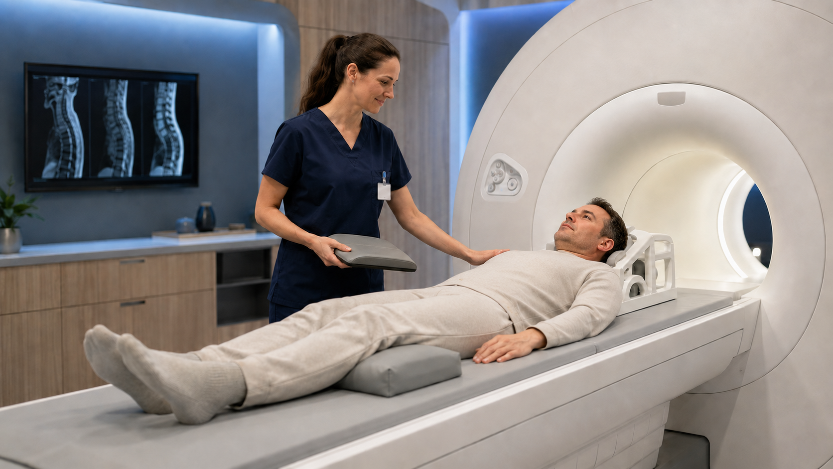
You will lie on a motorised table that moves into the MRI scanner. The scanner makes loud tapping and buzzing sounds, so you will receive hearing protection. The radiographer can see, hear and speak to you throughout the examination.
You will need to remain still because movement can reduce image quality.
At VitalScan, a whole spine MRI usually takes approximately 30 minutes. The exact time can vary depending on the images required and whether contrast is used.
What happens after a full spine MRI?
A radiologist will review the images and prepare a written report describing any relevant findings and whether further assessment is recommended.
At VitalScan, your radiologist report and digital images are normally available within three to five working days. They can be shared with your GP or specialist, who can interpret the findings alongside your symptoms and advise what should happen next.
How much does a private full spine MRI cost?
The cost of a private MRI scan depends on the regions examined, whether contrast is needed and what the appointment includes. When comparing private MRI scan costs, remember that a whole spine examination covers three regions rather than one.
VitalScan publishes its current price and inclusions clearly, so you can understand what you are paying for before you book. Check the latest full spine MRI cost and package details.
What can a full spine MRI not tell you?
MRI cannot always identify the precise cause of pain or prove that every visible change is responsible for your symptoms.
It also cannot replace a physical or neurological examination, confirm every infection or tumour without further testing, or assess every organ outside the spine.
This is why an MRI result should form one part of a wider clinical assessment.
When should you seek urgent medical help?
A routine private MRI appointment is not appropriate for symptoms suggesting serious spinal cord or nerve compression.
Seek urgent medical assessment if you develop:
- New loss of bladder or bowel control
- Difficulty starting or controlling urination
- Numbness around the genitals, buttocks or inner thighs
- Rapidly worsening weakness in one or both legs
- New difficulty walking or maintaining balance
- Severe back pain with significant neurological changes
- New spinal symptoms alongside a known history of cancer
- Severe pain following a major accident or injury
These symptoms do not always mean a serious condition is present, but they require prompt assessment. Do not wait for a routine scan appointment.
Why choose VitalScan for a private full spine MRI?
VitalScan provides private whole spine MRI scans at its Milton Keynes clinic, with:
- No GP referral required
- Cervical, thoracic and lumbar spine imaging in one examination
- Clinician advice included
- A full radiologist report
- Access to your digital scan images
- Results within three to five working days
- Free and accessible parking
- Support before and after your scan
Previous imaging, a referral letter or relevant medical information can still be helpful because it gives the team more context about the question that needs answering.
Not sure which MRI scan you need?
Choosing the correct scan matters. If your symptoms are limited to one area, a targeted MRI may be more appropriate than a full spine examination.
Contact the VitalScan team or call 01908 755556 before booking. We can explain the available options, although personalised medical advice may need to come from your GP or an appropriate specialist.
Ready to arrange your scan?
If you already know which examination you need, book your private MRI scan online. You can also view all private MRI scans available at VitalScan.
No GP referral required. Same-day and next-day appointments available in Milton Keynes. Our clinical team reviews every request to ensure the most appropriate scan is selected for your needs.
This article provides general information and does not replace personalised medical advice, diagnosis or emergency care