

Shoulder pain can make surprisingly simple movements difficult. Reaching for something on a shelf, putting on a jacket, lifting a bag or even sleeping on one side can become uncomfortable.
One possible cause is a rotator cuff injury.
The rotator cuff is a group of muscles and tendons that help stabilise your shoulder and allow you to lift and rotate your arm. These tissues can become irritated, strained or torn following an injury, repetitive movement or gradual wear over time.
However, shoulder pain does not always mean you have a rotator cuff tear. Bursitis, tendon irritation, arthritis, frozen shoulder and other problems around the shoulder can produce similar symptoms.
When the cause is unclear, a shoulder MRI scan can provide detailed images of the tendons, muscles and other structures inside the joint, helping your healthcare professional understand what may be contributing to your symptoms.
What Is a Rotator Cuff Injury?
Your shoulder is designed to move through a wide range of positions. That flexibility relies partly on four muscles and their tendons, collectively known as the rotator cuff.
The rotator cuff helps keep the upper arm bone positioned within the shoulder socket while you move your arm.
A rotator cuff injury can include tendon irritation or degeneration, inflammation, a partial-thickness tear or a full-thickness rotator cuff tear.
Some injuries happen suddenly. Falling onto an outstretched hand, lifting something heavy or experiencing a sports injury can damage the tendons.
Others develop more gradually. Repeated overhead movement, physical work, swimming, racquet sports, weight training or age-related changes in the tendons can contribute over time.
Importantly, structural changes in the rotator cuff do not always cause symptoms. Scan findings therefore need to be considered alongside your symptoms, movement and clinical assessment.
What Are the Common Rotator Cuff Symptoms?
Rotator cuff symptoms can vary depending on which tendon is affected and whether the problem developed gradually or followed an injury.
Pain is commonly felt around the outer or front part of the shoulder and may extend into the upper arm.
You may notice pain when:
- lifting your arm above shoulder height
- reaching behind your back
- putting on a coat or other clothing
- carrying or lifting objects
- exercising or performing overhead movements
- lying on the affected shoulder
Night pain can also occur, particularly when lying on the painful side.
Some people experience weakness when lifting or rotating their arm, reduced movement or clicking around the shoulder.
These symptoms cannot confirm a rotator cuff tear on their own because several shoulder conditions can produce a similar pattern.
Where Is Rotator Cuff Injury Pain Usually Located?
People searching for the rotator cuff injury location of pain often expect there to be one exact point.
In reality, the pattern varies.
Pain may be felt around the front or outer side of the shoulder and sometimes travels into the upper arm. Certain movements, particularly raising the arm away from the body or above the head, may make it more noticeable.
Pain beginning around the neck and travelling further down the arm, particularly alongside numbness or tingling, may indicate that the symptoms are not coming solely from the shoulder.
If you are unsure which area needs investigating, VitalScan’s guide MRI Scan for What? Start With the Question You Need Answered explains why understanding the pattern of your symptoms matters when choosing the right scan.
Rotator Cuff Tear or Something Else?
Shoulder conditions can overlap considerably.
A rotator cuff problem may cause pain and weakness. Frozen shoulder is more commonly associated with significant stiffness and restricted movement.
Bursitis can cause pain when lifting your arm, while arthritis may contribute to stiffness, aching and reduced movement.
Problems involving the shoulder labrum, biceps tendon or ligaments can also cause pain, clicking or a feeling that the shoulder is unstable.
Symptoms alone cannot always tell these conditions apart.
This is why ongoing or unexplained shoulder pain may need further assessment rather than trying to diagnose the problem based on symptoms alone.
When Might a Shoulder MRI Be Considered?
Not every painful shoulder requires an MRI.
A healthcare professional may first assess:
- how the pain started
- where the pain is located
- which movements trigger your symptoms
- whether there is weakness
- whether movement is restricted
- whether the problem followed an injury
- whether previous treatment has helped
Depending on the clinical question, an X-ray or ultrasound may also be considered.
A shoulder MRI may be useful when shoulder pain continues, significant weakness develops after an injury, the cause remains uncertain or detailed information about the soft tissues is required.
MRI can also be helpful when several structures inside the shoulder need to be assessed together.
VitalScan offers a dedicated Shoulder MRI Scan in Milton Keynes to assess the rotator cuff and surrounding structures.
You can also explore VitalScan’s wider range of private MRI scans if another part of the body needs investigating.
What Does a Shoulder MRI Show?
One of the main advantages of MRI is the amount of soft-tissue detail it provides.
Unlike an X-ray, which is particularly useful for assessing bones, MRI can examine the tendons, muscles, cartilage, ligaments and other soft tissues surrounding the shoulder.
Rotator Cuff Tendons
A shoulder MRI scan can provide detailed images of the tendons forming the rotator cuff.
It may identify changes such as:
- rotator cuff tendinopathy
- tendon degeneration
- partial-thickness rotator cuff tears
- full-thickness rotator cuff tears
- inflammation around the tendons
- tendon retraction in more substantial tears
This information can help your healthcare professional understand the location and extent of a rotator cuff injury.
Shoulder Muscles
MRI also allows the muscles connected to the rotator cuff to be assessed.
This can be particularly useful when a tendon injury has been present for some time, as longstanding tendon damage may be accompanied by changes within the surrounding muscles.
Bursa and Inflammation
The shoulder contains a small fluid-filled structure called the bursa, which helps reduce friction during movement.
Inflammation around the bursa can contribute to shoulder pain and may sometimes occur alongside rotator cuff problems.
MRI can provide information about fluid and inflammation around this area.
Shoulder Labrum
The labrum is a ring of cartilage surrounding the shoulder socket.
Although this article focuses on rotator cuff injuries, a shoulder MRI can also assess the labrum when another cause of pain, clicking or instability is suspected.
For some specific labral injuries, a specialist may recommend an Arthrogram MRI Scan, where contrast is introduced into the joint before imaging.
An MR arthrogram is different from a standard shoulder MRI and is generally used when additional detail is required for a particular clinical question.
Biceps Tendon and Ligaments
The long head of the biceps tendon passes through the shoulder and can sometimes contribute to pain.
MRI can examine this tendon as well as the ligaments and supporting soft tissues around the shoulder joint.
Bones, Cartilage and the Shoulder Joint
A shoulder MRI does not look only at the rotator cuff.
It can also provide information about the:
- shoulder bones
- cartilage
- joint surfaces
- surrounding soft tissues
- joint fluid
- other structural changes
This wider view can be useful when your symptoms could have more than one possible cause.
Can an MRI Show a Rotator Cuff Tear?
Yes. MRI can provide detailed images of the rotator cuff and may identify both partial-thickness and full-thickness tendon tears.
It can also provide information about the size and position of a tear and the condition of the surrounding muscles and tendons.
However, there is an important difference between finding a rotator cuff tear and proving that it is responsible for someone’s pain.
Rotator cuff changes can sometimes be present without causing symptoms, particularly as we get older.
Your MRI findings therefore need to be considered alongside your symptoms, examination and medical history.
The scan provides detailed information, but it is one part of the wider clinical picture.
Can an X-Ray Show a Rotator Cuff Tear?
A standard X-ray does not directly show a rotator cuff tendon tear because tendons are soft tissues.
X-rays can still be useful when investigating shoulder pain because they can show fractures, arthritis and other changes involving the bones and joint.
When the concern involves the rotator cuff itself, an ultrasound or MRI may provide more relevant soft-tissue information.
Shoulder MRI or Ultrasound: Which Is Better for a Rotator Cuff Injury?
Both ultrasound and MRI can play a role in investigating rotator cuff problems.
A shoulder ultrasound provides real-time imaging and can be useful for examining the rotator cuff tendons and bursa.
MRI provides a broader view of the entire shoulder and can assess the tendons alongside the muscles, labrum, cartilage, ligaments, bones and deeper structures.
The right test depends on what your healthcare professional is trying to investigate.
For a straightforward question about a tendon, ultrasound may sometimes provide the information required.
If symptoms are more complex, several structures need to be assessed or a wider view of the shoulder is needed, a shoulder MRI scan may be considered.
Can a Shoulder MRI Help After a Sports Injury
Shoulder injuries are common in sports involving repetitive overhead movement, impact or heavy loading.
This can include:
- swimming
- tennis and other racquet sports
- gym and weight training
- throwing sports
- contact sports
Following a sports injury, pain may not necessarily come from the rotator cuff alone.
The labrum, ligaments, biceps tendon, cartilage or other soft tissues can also be affected.
If pain, weakness or restricted movement continues following an injury, a shoulder MRI scan can provide detailed images of these structures and help investigate what may be causing the symptoms.
What Happens During a Shoulder MRI?
MRI is non-invasive and does not use ionising radiation.
Before your scan, you will complete an MRI safety assessment. It is important to tell the clinical team about implanted medical devices, previous operations, metal fragments or anything else that may affect MRI safety.
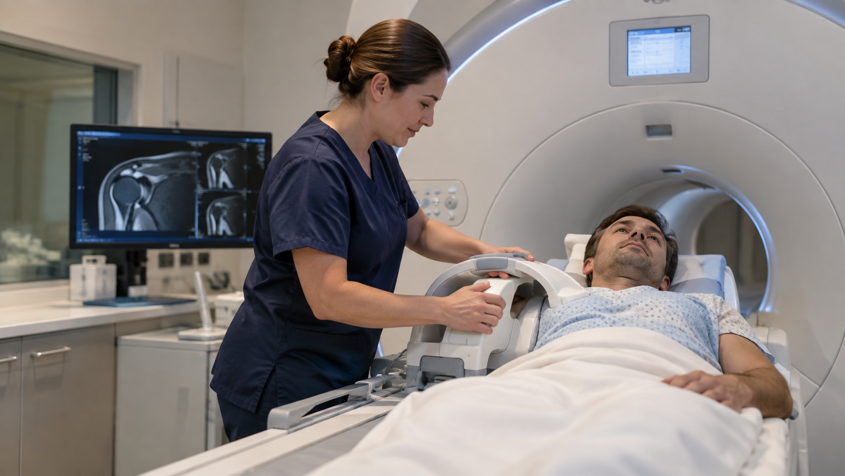
During the examination, you will lie on the scanning bed with your shoulder carefully positioned so detailed images can be captured.
The scanner makes tapping and knocking sounds while the images are being produced, so hearing protection is provided.
You will need to remain as still as possible during each imaging sequence to help produce clear pictures.
A shoulder MRI at VitalScan typically takes around 20 to 30 minutes, although the exact time can vary depending on the examination required.
If this is your first MRI, you can read VitalScan’s guide on How to Prepare for an MRI Scan before your appointment.
Does a Shoulder MRI Need Contrast?
Most routine shoulder MRI scans do not require contrast.
However, certain clinical questions involving structures such as the shoulder labrum or joint instability may require a different examination known as an MR arthrogram.
During an arthrogram, contrast is introduced into the joint before MRI imaging to provide additional detail.
A standard shoulder MRI and a shoulder MRI with arthrogram are therefore different examinations.
If an arthrogram has been recommended, you can find more information about VitalScan’s Arthrogram MRI Scan.
How Long Does a Shoulder MRI Take?
A shoulder MRI at VitalScan generally takes around 20 to 30 minutes.
During this time, you will need to keep your shoulder relatively still while the scanner captures a series of detailed images.
There is normally no recovery period following a standard MRI, so you can usually continue with your day afterwards.
What Happens After a Shoulder MRI?
Your MRI images are reviewed by a radiologist, who prepares a diagnostic report.
VitalScan’s shoulder MRI service provides a radiologist report and access to your digital scan images, with results typically returned within 3 to 5 working days.
You can share the report and images with your GP, physiotherapist, consultant or another healthcare professional involved in your care.
The MRI result alone does not determine whether you need physiotherapy, injections, surgery or another form of treatment.
Treatment decisions depend on several factors, including your symptoms, movement, strength, examination findings and the significance of anything seen on the scan.
When Should Shoulder Pain Be Assessed Urgently?
Most shoulder pain is not an emergency, but some symptoms should not simply be monitored while waiting for a routine scan.
Seek urgent medical assessment if you experience:
- severe pain following an injury
- inability to move your arm
- significant swelling
- an obvious change in the shape of the shoulder
- persistent numbness or loss of sensation
- an arm that becomes unusually hot or cold
- shoulder pain alongside fever or feeling seriously unwell
These symptoms may require urgent medical assessment rather than waiting for a routine MRI appointment.
Still Unsure What Is Causing Your Shoulder Pain?
Persistent shoulder pain can have more than one possible cause.
If you are experiencing ongoing pain, weakness, restricted movement or symptoms following an injury, imaging may help provide more information about what is happening inside the joint.
VitalScan’s Shoulder MRI Scan provides detailed imaging of the rotator cuff and surrounding shoulder structures at our Milton Keynes clinic.
If you are unsure whether a shoulder MRI is the right examination for your symptoms, read MRI Scan for What? Start With the Question You Need Answered or speak to the VitalScan team before booking.
Ready to take the next step? Book your MRI scan at VitalScan.
No GP referral required. Same-day and next-day appointments available in Milton Keynes. Our clinical team reviews every request to ensure the most appropriate scan is selected for your needs.